Alternate header for print version
Advanced search
Contributors
Help
Submit
Search
menu
Cell Process
Cell Component
Cell Type
Organism
Microbial
Alzheimer's
Data Sets
Center for Research in Biological Systems
University of California, San Diego
9500 Gilman Drive
La Jolla, CA 92093-0608, USA
Voice
: (858) 534-0276
Fax
: (858) 534-7497
Email
: dorloff@ncmir.ucsd.edu
Search Results for
endoplasmic reticulum organization
(2513 results)
(Not the results you were expecting?)
(Comments)
CIL:35999
NCBI Organism Classification
Gerbillinae
Biological Process
secretory granule organization
Cellular Component
secretory granule
Figures 379 (upper) and 380 (lower) from Chapter 15 (Cytoplasmic Inclusions) of 'The Cell, 2nd Ed.' by Don W. Fawcett M.D. Freeze substitution preparations of gerbil parotid gland show condensing vac...
CIL:9971
NCBI Organism Classification
Rattus
Biological Process
constitutive secretory pathway
Cellular Component
Golgi apparatus
Normal Rat Kidney (NRK) cells grown in culture expressing Galactosyl Transferase-YFP (GalT-YFP) and p58-CFP. This file is the CFP time series demonstrating the organization of the early secretory path...
CIL:11378
NCBI Organism Classification
Leporidae
Biological Process
post-translational protein modification
Cellular Component
Golgi apparatus
Figure 210 from Chapter 6 (Golgi Apparatus) of 'The Cell, 2nd Ed.' by Don W. Fawcett M.D. Supranuclear region of a cell from rabbit epididymis. Epithelial cells of the epididymis have a large Golgi a...
CIL:10841
NCBI Organism Classification
Rattus
Biological Process
autophagy
Cellular Component
autophagic vacuole
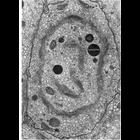
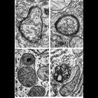
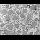
Figures 266 (upper left), 267 (upper right), 268 (lower left) and 269 (lower right) from Chapter 8 (Lysosomes) of 'The Cell, 2nd Ed.' by Don W. Fawcett M.D. Autophagic vacuoles from normal rat liver ...
CIL:35284
NCBI Organism Classification
Lilium formosanum
Biological Process
pollen tube growth
Cellular Component
mitochondria
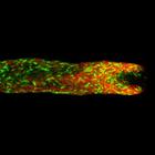
Living Lilium pollen tube labeled with mitotracker green to mark mitochondria (green) and cytochalasin D TRITC to mark endoplasmic reticulum (red) imaged using laser scanning focal microscopy. Shown i...
CIL:35289
NCBI Organism Classification
Lilium formosanum
Biological Process
pollen tube growth
Cellular Component
endoplasmic reticulum
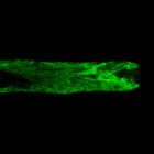
Living Lilium pollen tube labeled with mGFP5-HDEL to mark endoplasmic reticulum (green) imaged using laser scanning focal microscopy. Shown is a central 1.0 micron thick x-y slice. The growing tip is...
CIL:37236
NCBI Organism Classification
Cavia porcellus
Biological Process
protein secretion
Cellular Component
rough endoplasmic reticulum
Early transmission electron micrograph of secretory cell from the guinea pig pancreas. Secretory granules are prominent in the lumen of the rough endoplasmic reticulum. Image made available by James ...
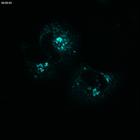
CIL:38939
NCBI Organism Classification
Mumps virus
Biological Process
response to virus
Cellular Component
endoplasmic reticulum
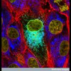
This confocal micrograph shows the mumps virus protein (turquoise) in the endoplasmic reticulum of a cultured cell. This is a region of the cell that processes proteins. This particular protein is pos...
CIL:40988
NCBI Organism Classification
Rattus rattus
Biological Process
protein synthesis
Cellular Component
microsome
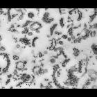
Transmission electron micrograph of a thin section of plastic-embedded material from the 'rough microsome' fraction of a preparation of rat liver. Subsequent to this early image, 'microsomes' became ...
CIL:40995
NCBI Organism Classification
Rattus rattus
Biological Process
protein synthesis
Cellular Component
rough endoplasmic reticulum
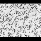
Transmission electron micrograph of a thin section of a preparation of isolated and plastic embedded rat liver polyribosomes (also known as polysomes). Polysomes associated with the endoplasmic reticu...
« Previous
1
...
11
12
13
14
15
16
17
18
...
252
Next »
Results per page:
10
20
50
100


")