Alternate header for print version
Advanced search
Contributors
Help
Submit
Search
menu
Cell Process
Cell Component
Cell Type
Organism
Microbial
Alzheimer's
Data Sets
Center for Research in Biological Systems
University of California, San Diego
9500 Gilman Drive
La Jolla, CA 92093-0608, USA
Voice
: (858) 534-0276
Fax
: (858) 534-7497
Email
: dorloff@ncmir.ucsd.edu
Search Results for
George E. Palade
(161 results)
(Not the results you were expecting?)
(Comments)
Still Images
Video/Animation
Z-Stack
Time Series
CIL:37161
NCBI Organism Classification
Rattus rattus
Biological Process
intestinal absorption
Cellular Component
microvillus
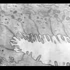
Transmission electron micrograph of rat intestinal colon showing apical microvilli. Image made available by James D. Jamieson and the Department of Cell Biology, Yale University School of Medicine.
CIL:37156
NCBI Organism Classification
Cavia porcellus
Biological Process
none specified
Cellular Component
none specified
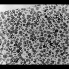
Transmission electron micrograph of a cell fractionation of golgi bottom fraction. Image made available by James D. Jamieson and the Department of Cell Biology, Yale University School of Medicine.
CIL:37141
NCBI Organism Classification
Rattus
Biological Process
exocytosis
Cellular Component
secretory granule
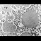
Transmission electron micrograph of exocytosis taken from the rat pancreas. Images such as these indicated that secretory granules closely and selectively approach the apical membrane of the pancreat...
CIL:7606
NCBI Organism Classification
Rattus rattus
Biological Process
biosynthetic process
Cellular Component
rough endoplasmic reticulum
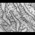
High magnification view of rough endoplasmic reticulum (RER) from rat pancreas showing RER and ribosomes, both bound (RiB) and free (RiF). Ribosomes were originally called Palade particles, as Palade ...
CIL:7598
NCBI Organism Classification
Cavia porcellus
Biological Process
canalicular bile acid transport
Cellular Component
tight junction
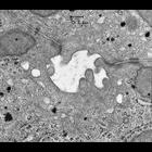
This is a view of apical domains of two adjacent hepatocytes showing the junctional complexes that attach cells to one another. The canaliculus is defined by the cell membranes of two adjacent liver c...
CIL:7599
NCBI Organism Classification
Cavia porcellus
Biological Process
canalicular bile acid transport
Cellular Component
tight junction
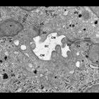
This is a view of apical domains of two adjacent hepatocytes in the guinea pig liver, showing the junctional complexes that attach cells to one another. The bile canaliculus (BC) is defined by the cel...
CIL:37188
NCBI Organism Classification
Rattus
Biological Process
glycogen metabolic process
Cellular Component
rough endoplasmic reticulum
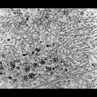
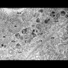
Transmission electron micrograph of liver cells from a 3-day old rat showing extensive smooth and rough endoplasmic reticulum, and glycogen (dark rosettes). Image made available by James D. Jamieson a...
CIL:7592
NCBI Organism Classification
Rattus
Biological Process
glycogen biosynthetic process
Cellular Component
glycogen granule
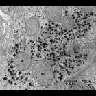
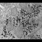
This section of liver from newborn rat shows glycogen (gly) deposits that are especially abundant in rat liver at this developmental stage. Deposits of glycogen, the storage form of glucose, are abund...
CIL:7591
NCBI Organism Classification
Rattus
Biological Process
glycogen biosynthetic process
Cellular Component
glycogen granule
This section of liver from newborn rat shows glycogen deposits that are especially abundant in rat liver at this developmental stage. Deposits of glycogen, the storage form of glucose, are abundant i...
CIL:7588
NCBI Organism Classification
Rattus
Biological Process
lipid digestion
Cellular Component
lysosome
This image shows the heterogeneity in the content of lysosomes (Lys) near the plasma membrane between two hepatocytes and near the bile canaliculus (not visible in this field). Some of the lysosomes ...
« Previous
1
...
7
8
9
10
11
12
13
14
...
17
Next »
Results per page:
10
20
50
100